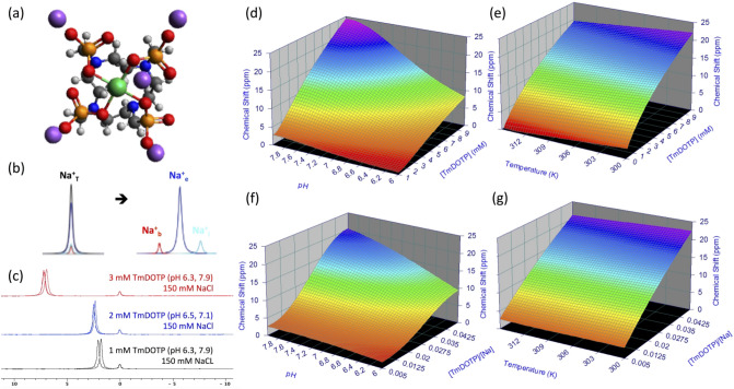
Figure 1.
Shifting mechanism of the 23Na resonance in vitro. (a) Chemical structure of sodium thulium(III) 1,4,7,10-tetraazacyclododecane-1,4,7,10-tetrakis(methylenephosphonate) (Na5TmDOTP). The TmDOTP5− complex consists of the Tm3+ ion chelated with DOTP8−. Each phosphonate-containing pendant arm on TmDOTP5- has electron-donating groups on the oxygen atoms (red) to stabilize the Tm3+ conjugation with DOTP8−. The -5 charge simultaneously attracts five Na+ ions (purple), which experience a shift in the observed 23Na resonance that is dependent on [TmDOTP5−]. (b) In vivo, prior to TmDOTP5− administration (left), the 23Na spectrum yields only a single peak representing the total sodium (Na+T) comprising blood (Na+b), extracellular (Na+e), and intracellular (Na+i ) compartments. Following TmDOTP5− administration (right), the peaks become spectroscopically separable based on [TmDOTP5−] in each compartment. Integrals of these peaks will be representative of aqueous [Na+] in each compartment. (c) A two-compartment coaxial cylinder tube setup was employed for in vitro observation of the chemical shift separation scheme (Figure S1). The inner tube (smaller volume) was filled with 150 mM NaCl, while the outer tube (larger volume) was filled with the same solution in addition to various amounts of TmDOTP5−, each subject to different pH conditions. Thus, all 23Na spectra from this phantom setup displayed a small unshifted peak from the inner compartment and a larger shifted peak. The outer-to-inner volume ratio was 8.6, explaining the difference in sizes of the peaks. Exemplary traces of 23Na spectra show that the shift is much more sensitive to [TmDOTP5−] (2.77 ppm/mM) than to variations in pH (0.25 ppm/pH unit) or temperature (0.03 ppm/°C). The downfield peaks in the red, blue, and/or black spectra are shifted differently due to varying TmDOTP5− concentrations, and these shifts exceed those caused by pH, but all of these shifts are detectable independent of broadening caused by TmDOTP5− (see also Figure S1). Plots (d,e) show that temperature, pH, and [TmDOTP5−] all contribute to variations of the 23Na chemical shift. However, these plots depict ranges of pH and temperature that are unlikely for in vivo settings (i.e., changes over 2 full pH units and temperature changes over 15 °C). Moreover, [Na+] in vivo (~ 150 mM in blood and extracellular space) is extremely high compared to [TmDOTP5−]. Therefore, variations in 23Na chemical shift are primarily dependent on [TmDOTP5−]/[Na+] thereby rendering (f) pH and (g) temperature dependencies negligible. Data points were fit to Chebyshev rational polynomials using TableCurve 3D v4.0.05 (Systat Software, San Jose, CA, USA; https://systatsoftware.com/products/tablecurve-3d/).

