Figure 7.
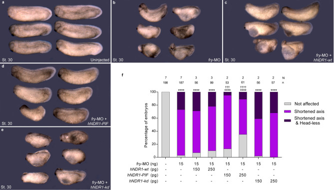
hNDR1-PIF partially rescues axis elongation in fry-depleted embryos. (a–e) 4-cell Xenopus embryos were injected into both dorsal blastomeres as indicated and fixed at St. 30. (a) Uninjected embryos. (b) fry-MO (15 ng) injected embryos. (c) fry-MO (15 ng) + hNDR1-wt mRNA (250 pg) co-injected embryos (d) fry-MO (15 ng) + hNDR1-PIF mRNA (250 pg) co-injected embryos. (e) fry-MO (15 ng) + hNDR1-kd mRNA (250 pg) co-injected embryos. Representative embryos are shown. (f) Quantitation of the percentage of embryos showing the different phenotypes: “Not affected”, “Shortened axis” or “Shortened axis & Head-less” phenotypes. Embryos were scored as “Shortened axis” when presented < 80% of the body length relative to the average length of control embryos. Data on graph is presented as mean. N: number of independent experiments, n: number of embryos. Statistical significance was evaluated using Chi-square test (****,††††p < 0.0001 and †††p < 0.001). * represents the comparison to the uninjected group and † represents the comparison to the fry-MO injected group.

