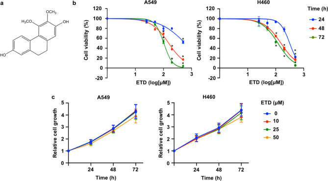
Figure 1.
Cytotoxicity of erianthridin (ETD) in A549 and H460 cells. (A) The chemical structure of ETD (3,4-dimethoxy-9,10-dihydrophenanthrene-2,7-diol) is shown. (B) A549 and H460 cells were treated with the indicated concentrations of ETD for 24, 48 and 72 h. Cell viability was analyzed using the MTT assay and is represented as a percentage. (C) A549 and H460 cells were treated with nontoxic concentrations of ETD for 24, 48 and 72 h. Cell proliferation was evaluated by the MTT assay. The rate of cell growth was calculated as a value relative to time 0 h. The data are presented as the mean ± SEM (n = 3). *p < 0.05 vs untreated control cells.

