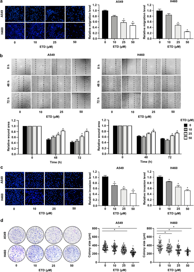
Figure 2.
ETD inhibits metastatic behaviors of non-small-cell lung cancer cells. (A) A549 and H460 cells were seeded onto a transwell chamber and treated with nontoxic concentrations of ETD (0–50 µM). After 20 h, the migrated cells were stained with DAPI and imaged by fluorescence microscopy. The scale bar is 10 µm. (B) A monolayer of the cells was scratched with a pipette tip to generate a wound, and the cells were treated with 0–50 µM ETD. The wound area was photographed under a microscope at 0, 48 and 72 h. The wound area was quantified at each time point relative to the area at the initial time point. (C) A549 and H460 cells were seeded onto a transwell chamber coated with Martigel and treated with nontoxic concentrations of ETD (0–50 µM). The scale bar is 10 µm. (D) Anchorage-independent growth assays were conducted by seeding cells into 24-well plates coated with 0.5% agarose. Cells were incubated with ETD and allowed to grow for 10 d. The colony size was measured using ImageJ54. Each dot plot represents a single colony. All data are presented as the mean ± SEM (n = 3). *p < 0.05 vs untreated control group.

