Figure 11.
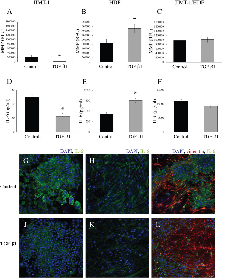
MMP activity and IL-6 level in the medium of JIMT-1 and HDF 3D mono-cultures and of JIMT1/HDF 3D co-cultures. The cultures were incubated in the absence (control) or presence of 5 ng/ml TGF-β1. After 14 days of incubation, the medium was collected for analysis of MMP activity (A–C) and the concentration of IL-6 (D–E). (A–C) The MMP activity was determined in 50 μl of medium by an assay based on the formation of a fluorescent product produced by MMP enzyme activity. (D–F) The level of IL-6 in the medium was determined by an ELISA assay. (G–L) Following the collection of the culture media, the cells were fixed with 3.7% formaldehyde and stained to visualize IL-6 (green), vimentin (red), and cell nuclei (blue). The images show a single confocal microscopy plane taken in the centre of the cultures. Each column represents mean ± SEM (n = 5). Students t-test, *P < 0.05. Scale bar is 50 μm.

