Figure 3.
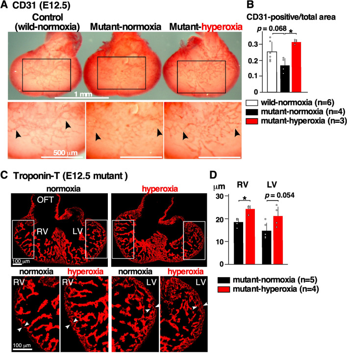
Defective coronary vessel organization in E12.5 Nkx2-5 mutant embryonic hearts, and the effects of hyperoxia. (A) Dorsal view of whole-mount CD31 immunostaining of ventricles showing endothelial cell plexus organized within the ventricles. CD31 staining was less prominent and extended to the apical area in Nkx2-5 mutant heart at normoxia, but was comparable at hyperoxia to the control wild-type heart (black arrowheads). (B) Quantitative CD31-positive area relative to the entire ventricular area. The number of embryos examined is indicated. The non-parametric Kruskal–Wallis ANOVA with Dunn’s test was used for analysis. (C) Representative heart sections stained with troponin-T. Thickness of compact layer is shown in white arrowheads. (D) Quantitative data showing thickness of compact layer in Nkx2-5 mutant hearts at normoxia vs. hyperoxia. The Mann–Whitney U test was used for analysis. Data are expressed as mean ± S.D.; individual data are represented as circles. *P ≤ 0.05. LV left ventricle, RV right ventricle, OFT outflow tract. The number of mice examined is indicated. ImageJ 1.52d (http://imagej.nih.gov/ij) was used for analysis.

