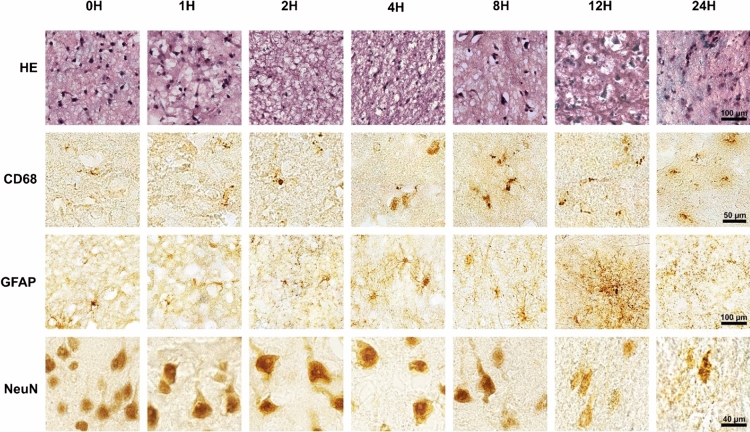
Figure 4.
Loss of neuronal staining progressed while micro- and astro-glial processes greatly expanded during the simulated PMI. From 0 to 2H, glia in the gray matter were mainly non-reactive and neurons (NeuN) were degrading, starting at 4H and peaking at 12H the activation of microglia (CD68) and astrocytes (GFAP) was seen with overlapping cellular processes. Small GFAP positive nodules appeared on astrocytic processes at 8H and increased so that by 12H they became the most prominent form of GFAP staining. At 24H, tissues appeared physically degraded with astrocyte cell bodies no longer identifiable, neurons (NeuN) mostly degraded, and rounded microglia. (HE = hematoxylin & eosin staining).

