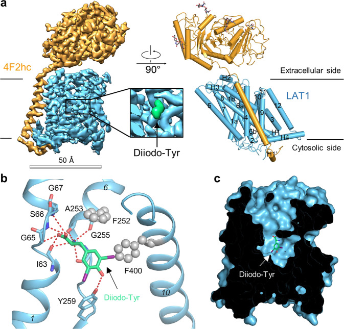
Fig. 3. Overall structure of the LAT1-4F2hc bound with Diiodo-Tyr.
a The overall structure of the LAT1-4F2hc bound with Diiodo-Tyr. The structure on the left is the EM map of the complex. The structure on the right is the overall structure of the complex. The glycosylation moieties are shown as sticks. H, helix. 4F2hc and LAT1 are colored orange and blue, respectively. b Diiodo-Tyr binding mode in LAT1. c LAT1 has a broad extracellular vestibule above Diiodo-Tyr.

