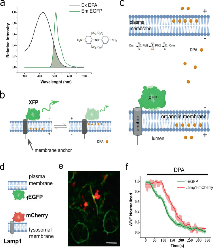
Fig. 1. Dipicrylamine (DPA) reaches internal membranes.
a Overlap between the spectra corresponding to EGFP emission and DPA absorption. A DPA molecule is pictured as inset. b Schematic representation of DPA transit in the HVoS technique. The negatively charged DPA accumulates preferentially at the positive leaflet of the membrane and redistributes in response to changes in membrane voltage. Redistribution of DPA within the membrane cause distance-dependent quenching of EGFP emission. c Schematic representation of DPA incorporation and distribution within the cell. Equilibrium describes the transit of DPA within a single membrane. Once at the membrane, DPA distributes according to a voltage-dependent equilibrium with voltage-dependent rates v1 and v2. d Cartoon depicting the topology of the probes used for simultaneous measurements of plasma membrane and lysosomes. e Simultaneous expression of Lamp1-mCherry (lysosomal marker) and farnesylated GFP (fEGFP; plasma membrane marker) in HEK293 cells. Bar = 10 μm) f Time course of fluorescence for the cell in panel e during DPA incorporation. The lysosomal signal (red trace) corresponds to the average of six spots proximal to the membrane and centered in the cell. The plasma membrane signal (green trace) was extracted from four regions of equal size.

