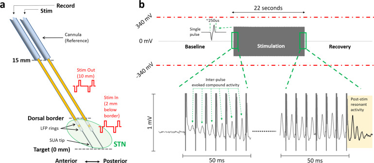
Fig. 1. Experimental paradigm and sample stimulation segment showing that LFP recordings did not saturate with the high input range of the amplifier.
a The microelectrode diagrams depicting the recording and stimulating electrodes. The “out-STN” stimulation was performed 10 mm above the ventral border of the STN. Bipolar microelectrodes with two 0.5-mm wide stainless-steel rings separated by 0.5 mm were used to deliver biphasic electrical stimulation and record LFP activity. The “in-STN” stimulation experiments were performed 2 mm below the dorsal border of the STN, characterized by the increased background activity and neuronal spiking recorded from the fine microelectrode tip. b Sample 66 s raw LFP recording illustrates that the amplifier was not saturated during stimulation of the other electrode. Single pulse waveform illustrates that the biphasic stimulation pulse was captured within a short time, allowing the LFP recordings to continue with minimal interruption. Zoomed 50 ms segments from beginning and the end shows evoked potentials induced with each stimulus pulse. The evoked waveform amplitude increases with each pulse and settles after ~10 pulses. With the termination of the stimulation, the resonance in the evoked activity can be observed longer, which dampens within 20 ms.

