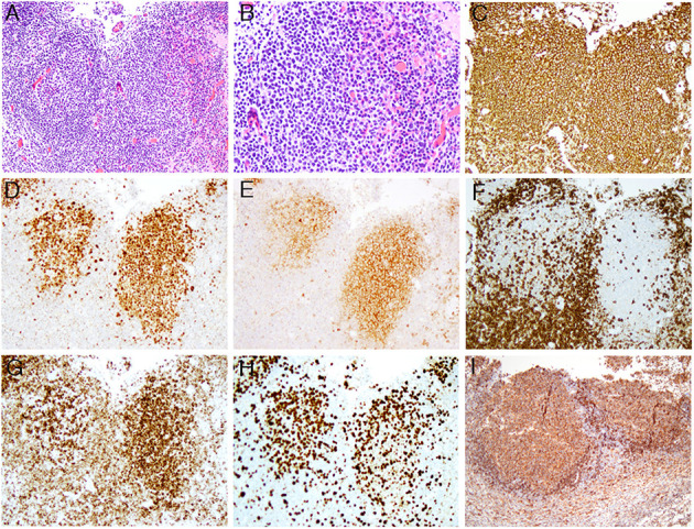
Figure 1.

The composite microphotographs of low-grade FL with high proliferation index. (A,B) The follicular lymphoma cells are mainly centrocytes with <5 centroblasts per high power field [the original magnifications of (A,B) are 100x and 200x, respectively]; (C–E) the lymphoma cells are positive for CD20 (C), BCL6 (D), and CD10 (E) [the original magnifications of (C–E) are 100x, 100x, and 100x, respectively]; (F–H) In comparison to negative CD3 (F), the lymphoma cells are positive for BCL2 (G) with high Ki-67 proliferation index (H) [the original magnifications for (F–H) are 100x, 100x, and 100x, respectively]; (I) The lymphoma cells are positive for HLA-DR (the original magnification is 100x).
