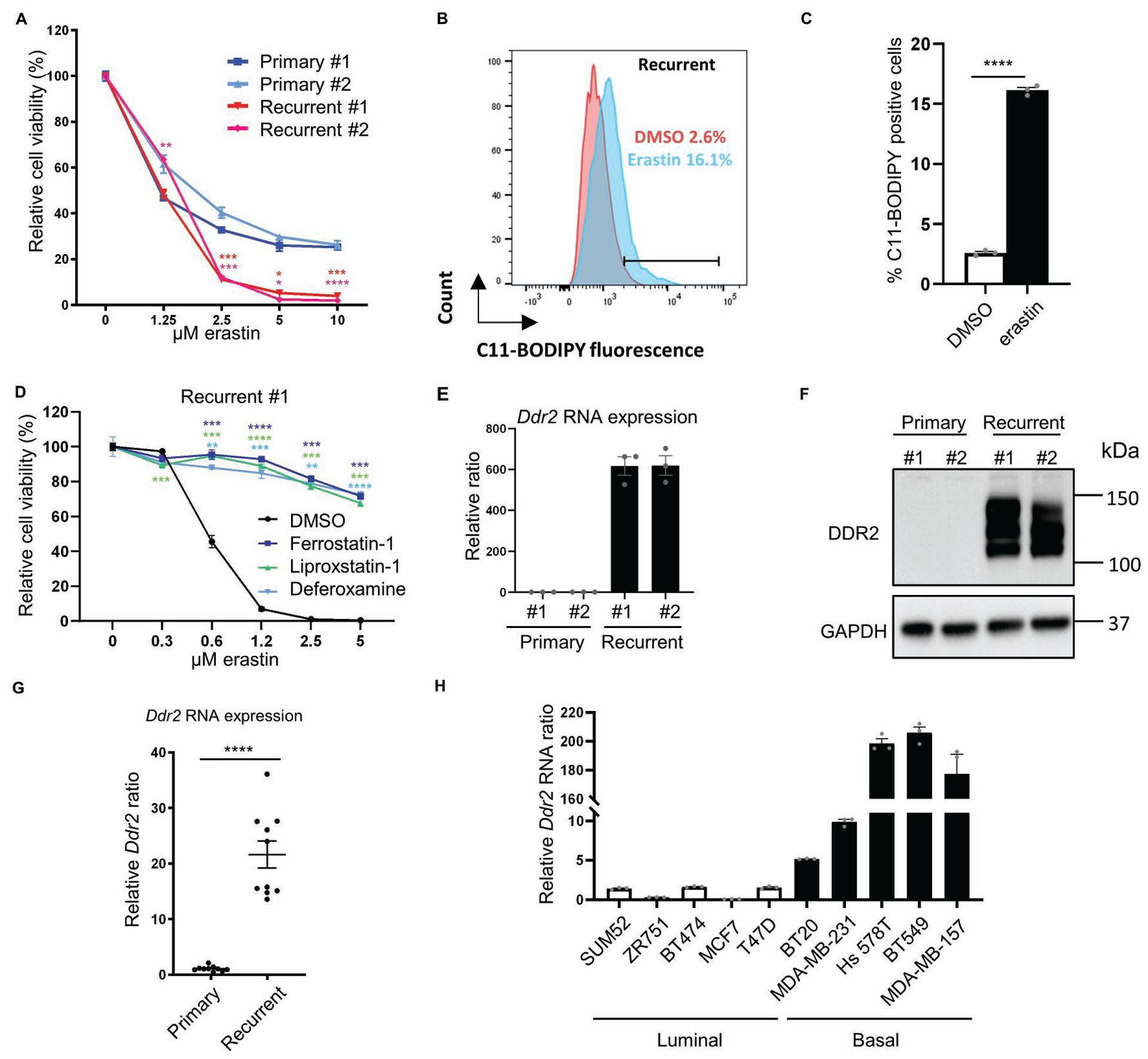
Fig. 1. Recurrent tumor cells are sensitive to ferroptosis and show high DDR2 expression.

(A) Recurrent tumor cells were more sensitive to erastin treatment. Primary and recurrent murine tumor cells were treated with increasing indicated doses of erastin for 18 hours and the viability was measured by Celltiter Glo assay. n=3 biological replicates. (B-C) Erastin treatment (0.5μM, 18 hours) in recurrent tumor cells increased lipid peroxidation as determined by C11-BODIPY staining (B) and the quantification of % positive cells (C). (D) The erastin-induced cell death in recurrent tumor cells was rescued by ferroptosis inhibitors (ferrostatin-1, 10 μM; liproxstatin-1, 2 μM) and iron chelator (deferoxamine, 100 μM) as determined by Celltiter Glo assay after 19 hours of incubation. n=3 biological replicates. (E) Ddr2 expression was dramatically elevated in recurrent tumor cells by qRT-PCR. (F) Western blot showed a robust DDR2 protein expression only in recurrent tumor cell lines. (G) Comparison of Ddr2 RNA expression by qRT-PCR between 10 primary and 10 recurrent mouse tumors showed an overall increase in recurrent tumors. (H) DDR2 RNA is highly expressed in human basal breast cancer cell lines. Comparison of DDR2 RNA expression between 5 luminal (empty bars) and 5 basal (solid bars) established human breast cell lines expressed higher levels of DDR2 mRNA. (A, D) Two-way ANOVA, *p < 0.05, **p < 0.01, ***p < 0.001, ****p < 0.0001 Dunnett’s multiple comparisons. (C, G) ****p<0.0001, two-tailed Student’s t-test. n = 10 samples for each group. Bars show standard error of the mean.
