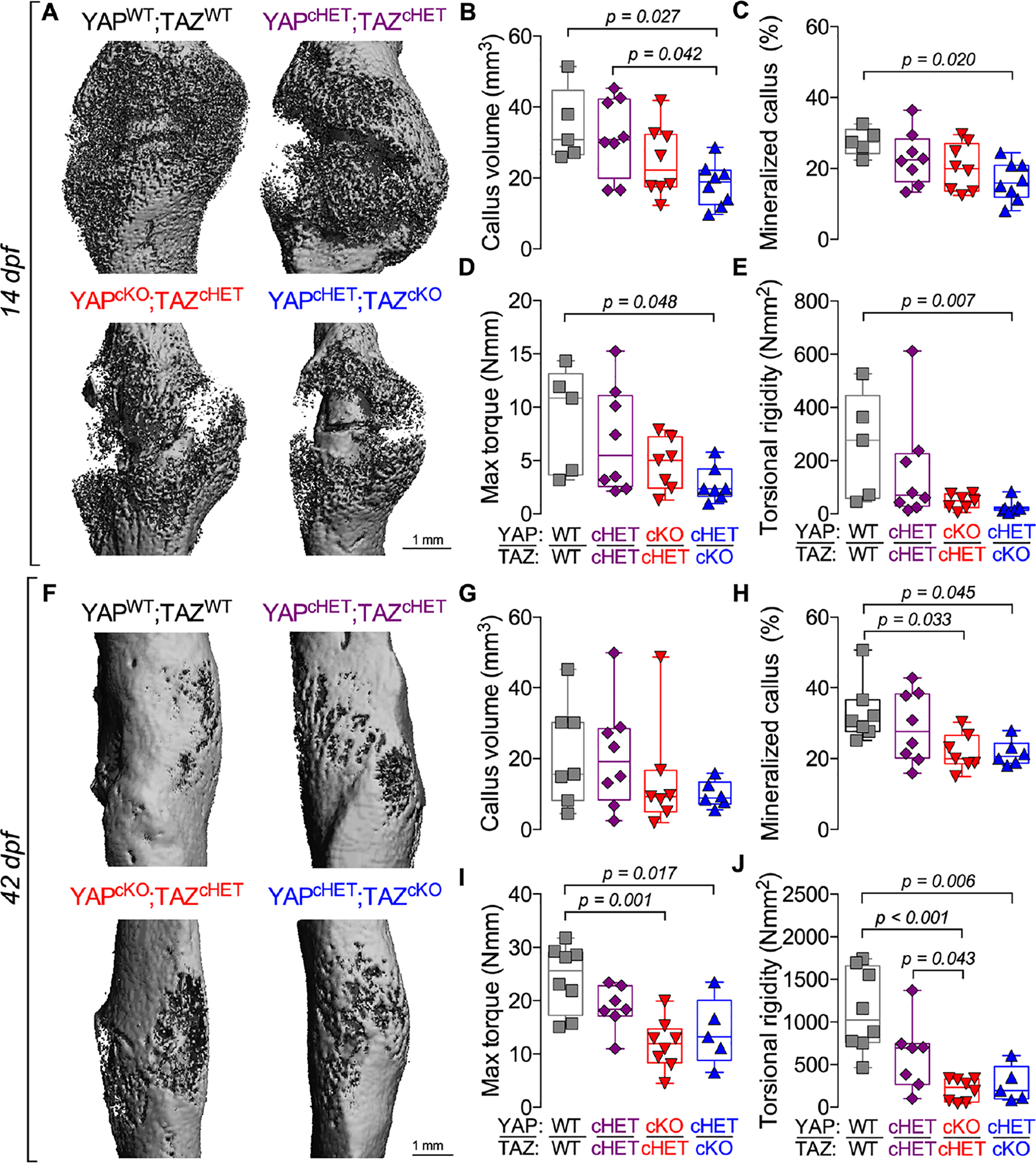
Figure 1. Constitutive, combinatorial YAP/TAZ deletion from Osterix-expressing cells impaired fracture healing.

A) MicroCT reconstructions at 14 days post-fracture (dpf). Quantification of 14 dpf callus architecture: (B) total callus volume and (C) mineralized callus percentage. Quantification of 14 dpf callus mechanical testing in torsion to failure: (D) maximum torque and (E) torsional rigidity. F) MicroCT reconstructions at 42 dpf. Quantification of 42 dpf callus architecture: (G) total callus volume and (H) mineralized callus percentage. Quantification of 42 dpf callus mechanical testing in torsion to failure: (I) maximum torque and (J) torsional rigidity. Data are presented as individual samples in scatterplots and boxplots corresponding to the median and interquartile range. Data were evaluated by one-way ANOVA with post-hoc Tukey’s multiple comparisons tests. Groups with significant pairwise comparisons are indicated by bracketed lines p-values, adjusted for multiple comparisons. Sample sizes, N = 5–8. Scale bars indicate 1 mm for microCT reconstructions.
