
Figure 8. Adult onset-inducible, homozygous YAP/TAZ deletion from Osterix-expressing cells impaired periosteal osteoblast precursor bone formation.
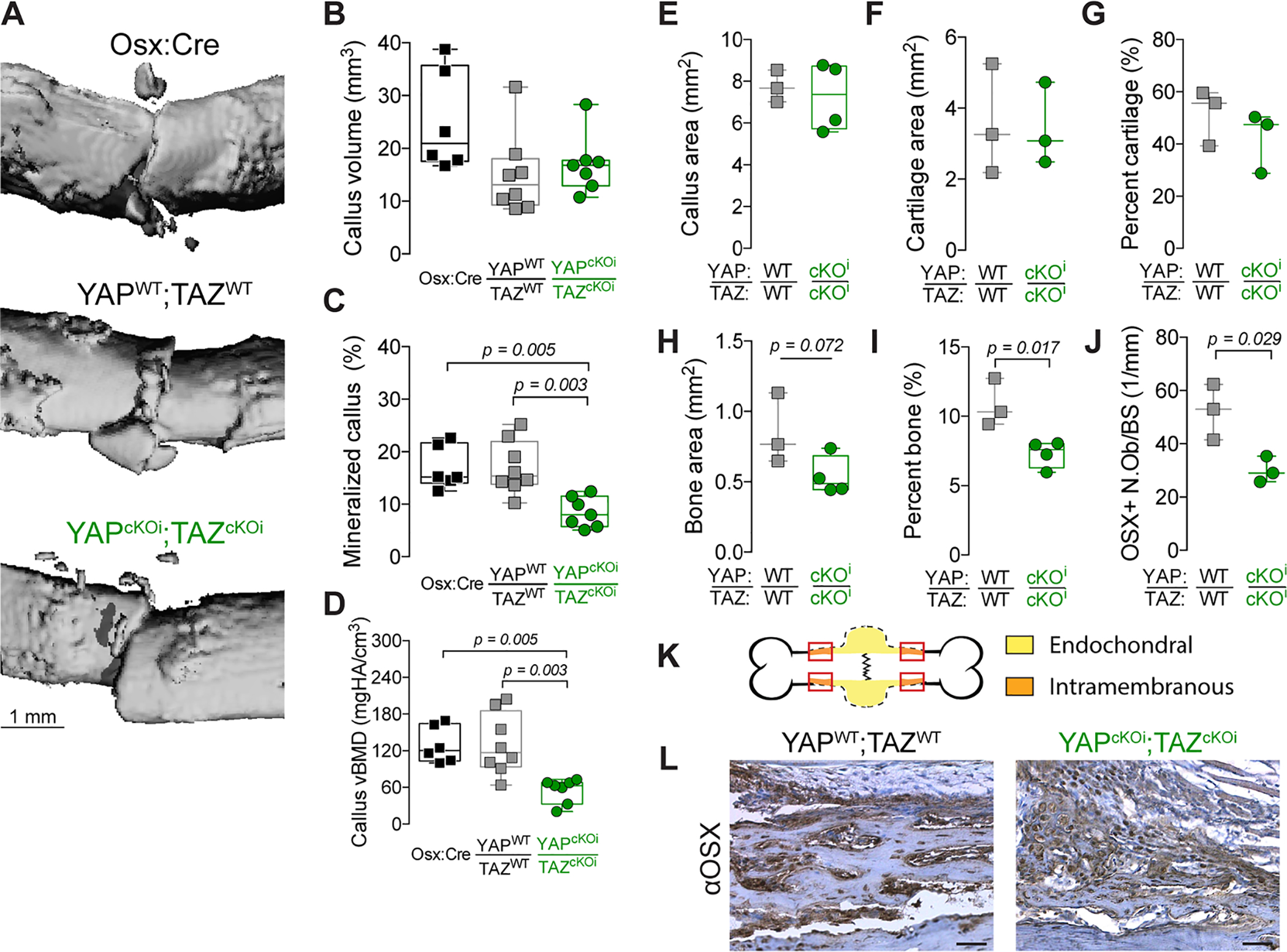
A) MicroCT reconstructions at 7 days post-fracture (dpf). Quantification of 7 dpf callus architecture: (B) callus volume, (C) mineralized callus percentage and (D) volumetric bone mineral density. Quantification of total callus histomorphometry at 7 dpf of (E) total callus area, (F) cartilage area, (G) percent cartilage, and (H) bone area, and (I) percent bone area, and (J) Osterix-positive osteoblasts per bone surface (OSX+ N.Ob/BS). K) Representation of the callus “shoulder,” where bone formation initiates by intramembranous ossification, and where we evaluated (L) anti-Osterix (αOSX) immunostaining at 7 dpf. Data are presented as individual samples in scatterplots and boxplots corresponding to the median and interquartile range. When appropriate, an independent t-test was used and p values are shown with bracketed lines. If not shown, p value for comparison > 0.05. Sample sizes, N = 3–8. Scale bars indicate 1 mm for microCT reconstructions and 50 μm for micrographs.