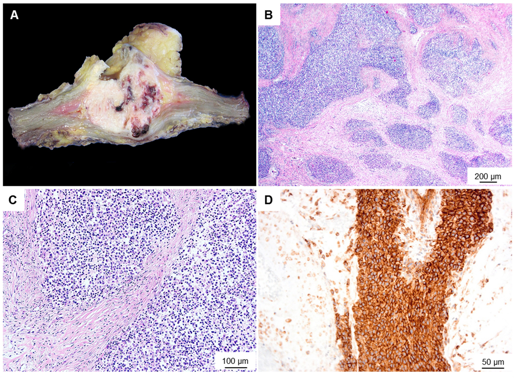
Figure 1. Macroscopy and microscopy images of the Case 1.

(A) Macroscopic image shows a fairly well circumscribed fleshy tumour with foci of necrosis and haemorrhage. (B) Microscopic image (low-power) shows the tumour to be composed of solid sheets and nests surrounded by a desmoplastic stroma. (C) High power view of the tumour shows small sized cells with scant cytoplasm. (D) Membranous staining with CD99- mimicking Ewing’s sarcoma.
