Figure 5.
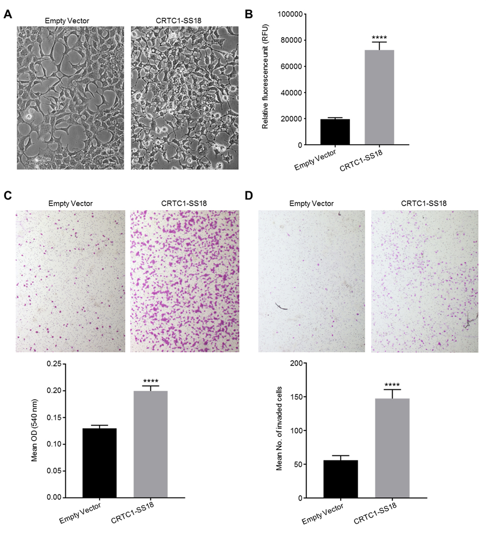
Biological analysis of the CRTC1-SS18 gene fusion. (A) Morphological analysis of CRTC1-SS18 expressing cells. CRTC1-SS18 fusion-expressing clones were morphologically distinct from control HEK293s; the cytoplasmic component was small, with pronounced vacuoles and extended, thin pseudopodia (both images taken under phase contrast at 200X magnification). (B) Expression of the CRTC1-SS18 fusion protein in HEK293 cells significantly increased anchorage-independent growth potential. The number of viable, colony-forming cells present, following incubation in soft agar was increased 2.1-fold in HEK293 cells expressing CRTC1-SS18 compared to HEK293 cells transfected with a control plasmid. Viable cells were determined by a fluorometric assay. p<0.0001, t=8.61, df=14. Error bars represent SEM (n=8). (C) Expression of the CRTC1-SS18 fusion protein in HEK293 cells significantly increased cell migration. The number of HEK293 cells expressing CRTC1-SS18 that migrated through 8 μm pores in a Boyden chamber assay in 16 h was significantly increased compared to HEK293 cells transfected with a control plasmid. This was apparent by inspection by microscopy (at 4X objective magnification) and by a colorimetric assay. p<0.0001, t=6.2.2, df=38. Error bars show SEM (n=20). (D) Expression of the CRTC1-SS18 fusion protein in HEK293 cells significantly increased cell invasive potential. The number of HEK293 cells expressing CRTC1-SS18 that invaded through an 8 μm pores coated in a basement membrane matrix in 16 h was significantly increased compared to HEK293 cells transfected with a control plasmid. Invaded cells were counted at 40X objective magnification, the mean number of invaded cells was 2.6-times greater for CRTC1-SS18 expressing cells than for control cells. p<0.0001, t=6.108, df=42. Error bars show SEM (n=22).

