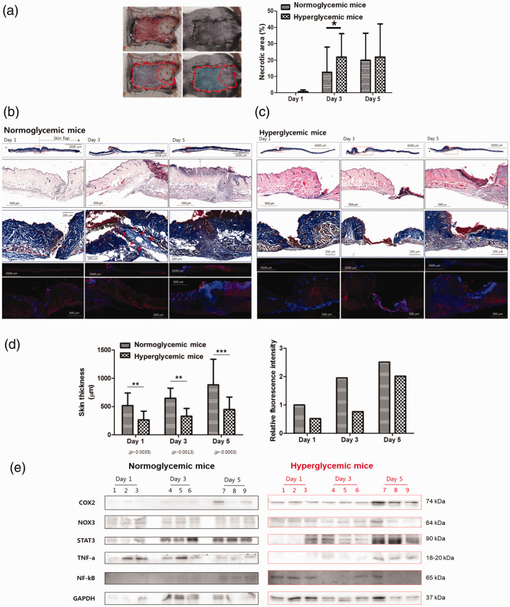
Figure 1.
Wound healing in normo- and hyperglycemic mice. (a) Full-thickness wounds were created on the backs of control and streptozotocin-induced hyperglycemic mice. The wounds were examined one, three, and five days after wound creation. The extent of the difference in the necrotic area (%) between normo- and hyperglycemic mice model. (b, c) H&E and CD31-stained representative sections of (b) normoglycemic mice and (c) hyperglycemic mice wound harvested one, three, and five days after wound creation. (d) The quantification of skin thickness (μM) and relative fluorescence intensity of normoglycemic and hyperglycemic mice one, three, and five days after wound creation. (e) The expression of COX2, NOX3, STAT3, TNF-α, and NF-κB was calculated in normoglycemic and hyperglycemic mice examined one, three, and five days after wound creation. Measurements were made in duplicate in three mice in each group and each day. Western blots are representative of three different experiments. Results are expressed as mean ± SD. (A color version of this figure is available in the online journal.)

