Figure 1.
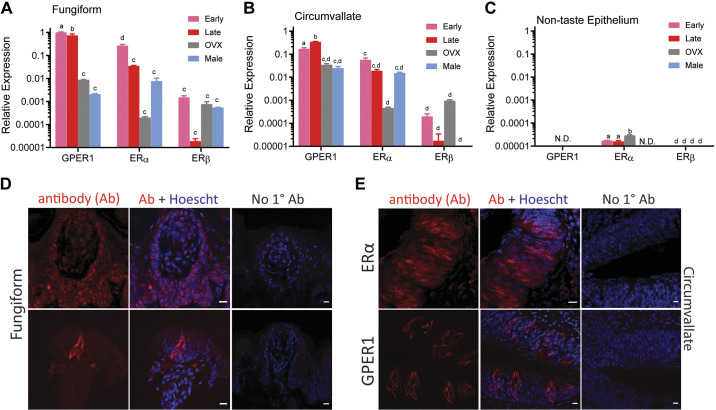
Estrogen receptor (ER) expression in mouse taste cells is altered during estrous. Quantitative real time PCR was used to determine relative expression of ERs in taste buds from the fungiform papillae (A), circumvallate papillae (B), and nontaste epithelium (C). Bars depict mean ± SE gene expression relative to the internal calibrator [early-phase G protein-coupled estrogen receptor 1 (GPER1) in fungiform papillae] of 3 independent biological replicates using GAPDH as the housekeeping gene. N.D. denotes gene was not detected in the qPCR assay. Letters (a,b,c,d) above bars indicate statistical grouping variables determined using 2-way ANOVA corrected for multiple comparisons using Tukey–Kramer method. D and E: confocal images illustrating ER localization in taste cells from fungiform and circumvallate papillae, respectively. ERα is found primarily as a nuclear receptor in taste cells and the nonclassical ER; GPER1 protein is localized in taste cells as membrane and cytosolic receptor. Scale bars, 10 µm.

