Figure 2.
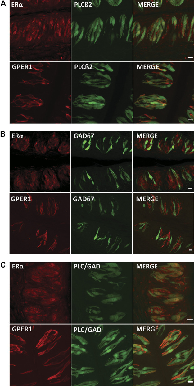
Estrogen receptors (ERs) are present in type II cells. A: representative immunofluorescent images for ERα and G protein-coupled estrogen receptor 1 (GPER1; red) in vallate taste buds from mice expressing GFP in phospholipase Cβ2 (PLCβ2)-positive (type II) cells, and the corresponding merged image. Yellow color indicates both ERs and PLCβ2 within the same taste cells. Note that some ER-positive cells are not colocalized with PLCβ2. Scale bars, 10 µm. B: immunofluorescence for ERs and a marker [GFP-glutamic acid decarboxylase 67 (GAD67)] for type III cells in mouse vallate taste buds. Scale bars, 10 µm. C: immunofluorescence assays for ERs in double-GFP transgenic mice show that there was little GPER1 receptor expression in the population of non-type II and non-type III vallate taste cells. ERα colocalize with GFP-positive cells and are apparent in some non-GAD67- or PLCβ2-expressing cells (green). Scale bars, 10 µm.

