Figure 4.
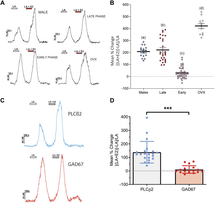
Acute exposure of estrogen increases taste cell responses to linoleic acid. A: Representative traces of the intracellular calcium ([Ca2+]i) change induced by 10 µM linoleic acid (LA) and LA with 17β-estradiol (E2, 10 nM) in taste cells of wild-type (WT) males, females during early phases, late phases, and ovariectomized (OVX) females. B: summary data showing mean percent change of responding taste cells comparing LA (10 µM) alone to the combination of LA and estradiol (10 nM). Individual data points represent one taste cell (WT males, n =16; females during estrous phase, n = 28; females during proestrus phase, n = 40; and ovariectomized females, n = 11). Different letters represent different statistical groups determined by one-way ANOVA with Tukey–Kramer method for post hoc multiple comparisons. C: enhancement of fatty acid responses by estrogen is found only in type II cells. Taste cell responses were recorded from isolated taste cells (n = 37) of female mice on estrous stage. Cells were sequentially stimulated with LA (10 µM) and LA + E2 (10 nM) in random order. Traces show responses from phospholipase Cβ2 (PLCβ2)-GFP cells (blue) and glutamic acid decarboxylase 67 (GAD67)-GFP cells (red). D: increased responsiveness to the combination of LA and E2 was observed primarily in cells expressing PLCβ2-GFP but not cells expressing GAD67-GFP. ***P < 0.001 as indicated by Student’s t test.

