Figure 1.
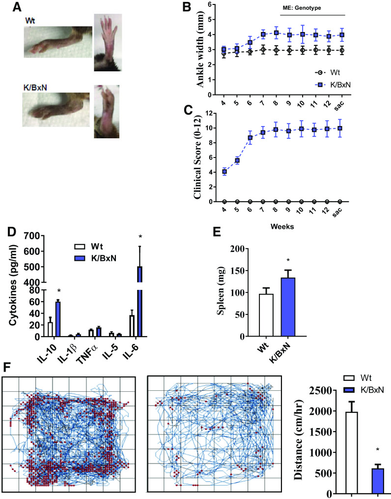
The K/BxN mouse model of RA shows joint pathology, systemic inflammation, and reductions in physical activity. A: representative image of hind ankle joints in wild-type (Wt) and K/BxN mice. B: time course of ankle width measurements taken throughout the 13-wk study design. C: joint clinical score for front and hind paws through the 13-wk study. (For B and C; n = 10 male mice/group; repeated-measures ANOVA, main effect of genotype, *P < 0.05). Plasma cytokine levels (D) and spleen weights (E) taken at 13 wk from wild-type and K/BxN mice. F: open-field activity measurements taken throughout 60 min in wild-type and K/BxN mice. Representative activity maps for each group (left). Quantification of distance traveled per hour (right). (For D–F; n = 10 male mice/group; two-tailed t test, *P < 0.05). Data are presented as means ± SE. RA, rheumatoid arthritis.

