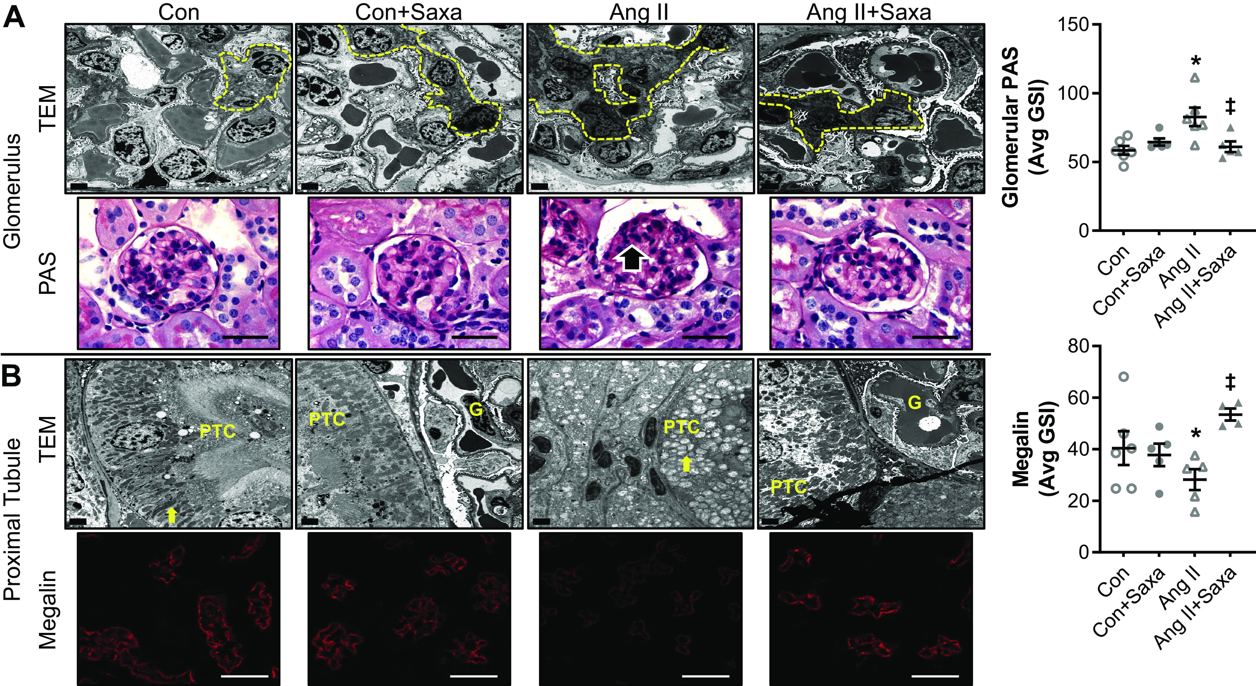
Figure 3.

ANG II-induced kidney cortical injury is attenuated by Saxa. Glomeruli of ANG II-infused mice demonstrated focal and segmental mesangial expansion (dashed yellow lines, black arrow), assessed by TEM and PAS staining (600×), that was attenuated by Saxa (A). Proximal tubules of ANG II-infused mice exhibited loss of microvillar architecture and basilar canaliculi and emergence of mitochondrial rounding, assessed by TEM, as well as reduced brush border megalin expression that were both attenuated by Saxa (B). Glomerular PAS staining and brush border megalin expression quantitation are shown in panels on the right. n = 4–6 mice per group. Statistical significance was determined using two-way ANOVA followed by Fisher’s LSD post hoc analysis. *P < 0.05 vs. all other groups, ‡P < 0.05 vs. ANG II. ANG II, angiotensin II; Con, control; G, glomerulus; PAS, periodic acid-Schiff; PTC, proximal tubule cell; Saxa, saxagliptin; TEM, transmission electron microscopy.
