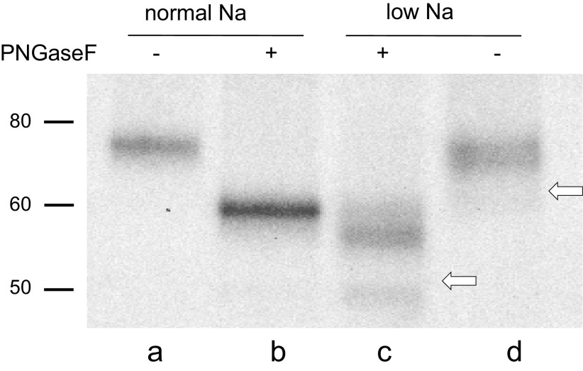
Figure 6.
Western blot of γENaC in microsomes of rat kidney before and after treatment with PNGaseF under nonreducing conditions. Rats were fed either control or low-Na diets. All lanes were loaded with material corresponding to 20 µg of microsomal protein. In rats on a control diet, the full-length form predominates. In these gels, this form migrates at 75 kDa before (lane a) and 60 kDa after (lane b) PNGaseF treatment. In rats on a low-Na diet, the doubly cleaved forms predominate (lanes c and d). These migrate as a broad band at 70 kDa before and at ∼56 kDa after PNGaseF. The white arrows show the locations expected of the fully cleaved species under reducing conditions. The positions of the cleaved forms are consistent with excision of 3 to 4 kDa from the protein. The blot is representative of three different experiments. ENaC, epithelial Na channel.

