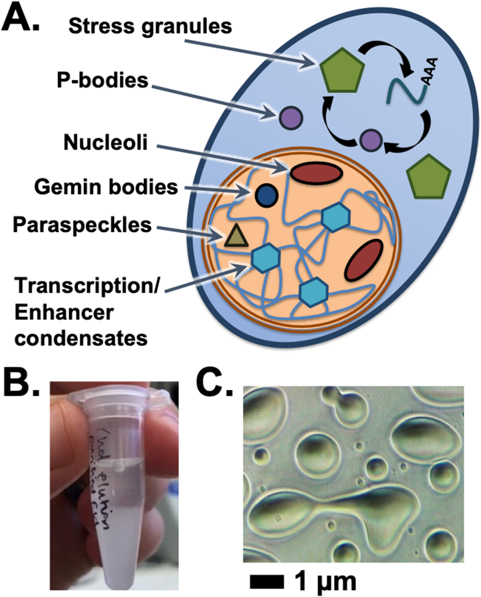
Figure 1.

Condensates in cells and in vitro. (A) The cell has a number of granule bodies and non-membrane bound organelles, including those listed here in the cytoplasm or nucleus. (B) Phase separation can be visually observed by the accumulation of turbidity and eventually separation of protein condensate by gravity at sufficiently high protein concentrations. (C) Microscopic scale condensates allow the fluid and dynamic properties of protein phase separation to appear as condensates assume droplet shapes, fuse together, and wet surfaces on which they settle.
