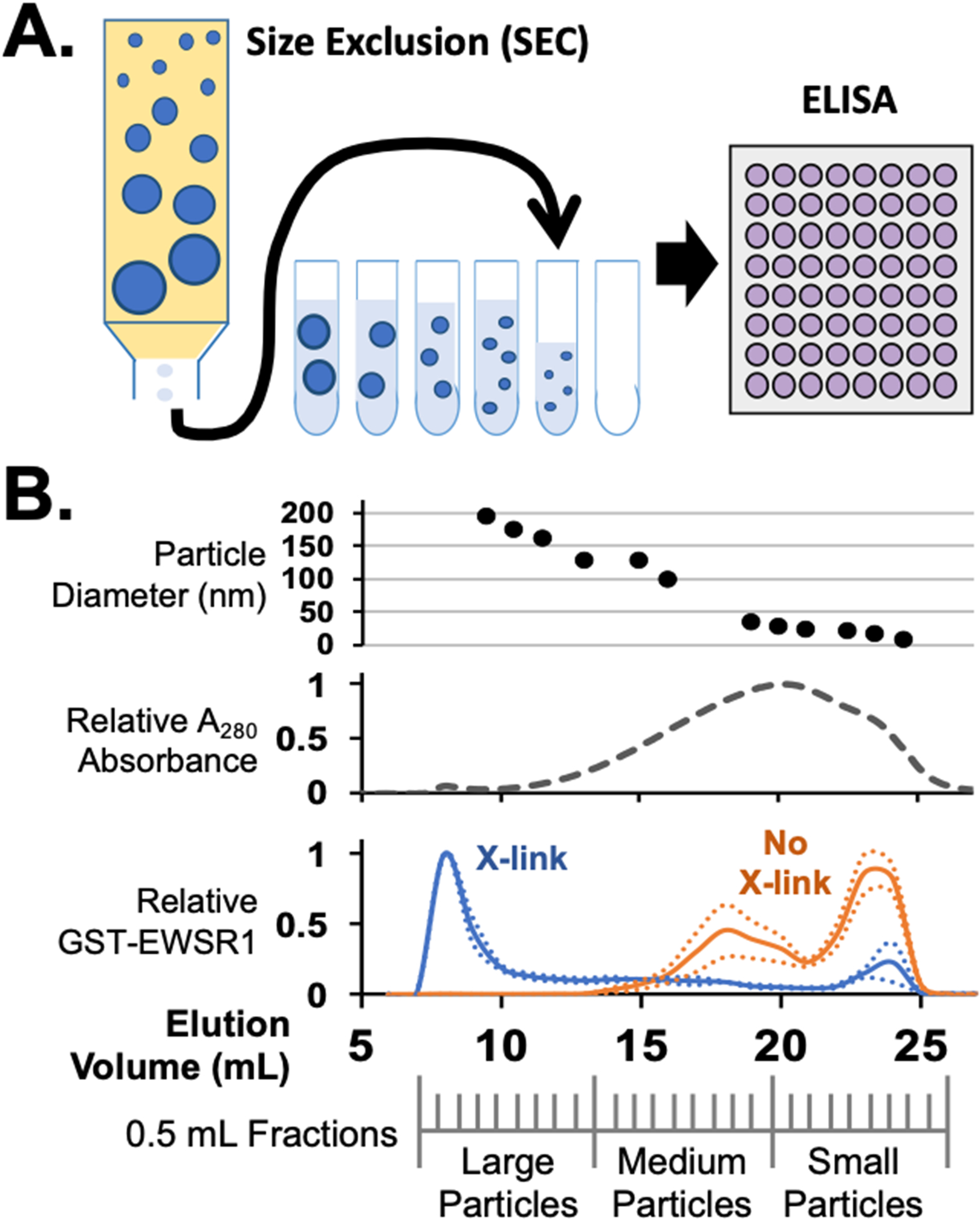
Figure 2.

Separation of cell condensates by size exclusion chromatography. (A) Cell assemblies and condensates stabilized by formaldehyde crosslinking can be separated by SEC. ELISA assays of fractions collected can reveal the elution profile for specific crosslinked proteins that elute. (B) Example of SEC data from E. coli lysates expressing the protein GST-EWSR1. Particle sizes eluted can be measured by either DLS or TEM. Shown here is DLS data indicating particles between 200 and 10 nm measured between early and late fractions respectively, top. By monitoring UV absorption, most crosslinked proteins in a cell lysate elute in late fractions as small complexes or monomers. Protein eluting from SEC of a no-crosslink cell lysate produce the same UV absorption profile. In an ELISA, GST-EWSR1 was found to elute primarily in early fractions as large particles, blue. Without crosslinking, GST-EWSR1 assemblies break apart and ELISA signals are primarily in the late fractions, orange, consistent with medium to small particles.
