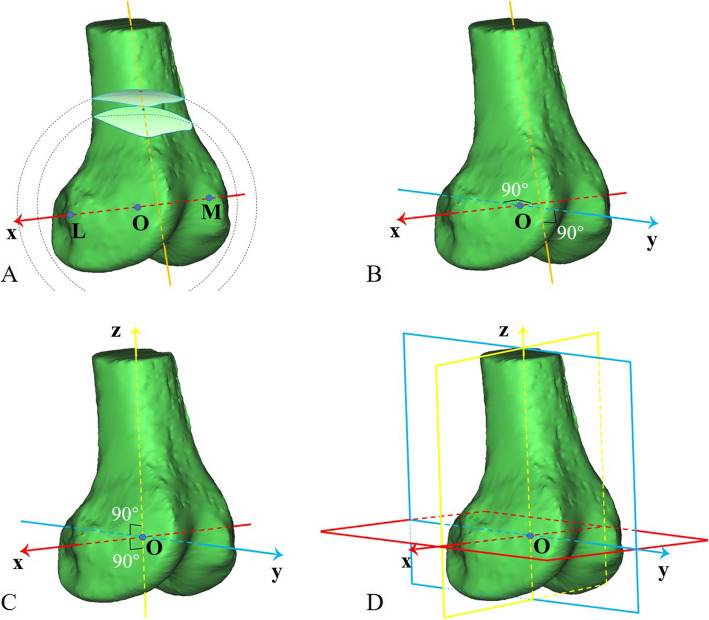
Fig. 2.
A coordinate system was established based on the femur. a The fovea of the medial epicondyle (point M) and the highest point of the lateral epicondyle (point L) were selected to form the TEA, which was defined as the x-axis, with the midpoint of the TEA as the origin (point O); two virtual balls—with the origin as their centres and with 0.7 and 0.8 times of the TEA length as their radius respectively—were crossed with the femur to obtain two section surfaces, whose centroids were linked to determine the femoral shaft axis. b The y-axis was defined as the line passing through the origin and perpendicular to the TEA and the femoral shaft axis meanwhile. c The z-axis was perpendicular to the x-axis and y-axis through the origin. d Three planes were determined by the x-, y- and z-axes (red: level plane; yellow: coronal plane; blue: midsagittal plane)

