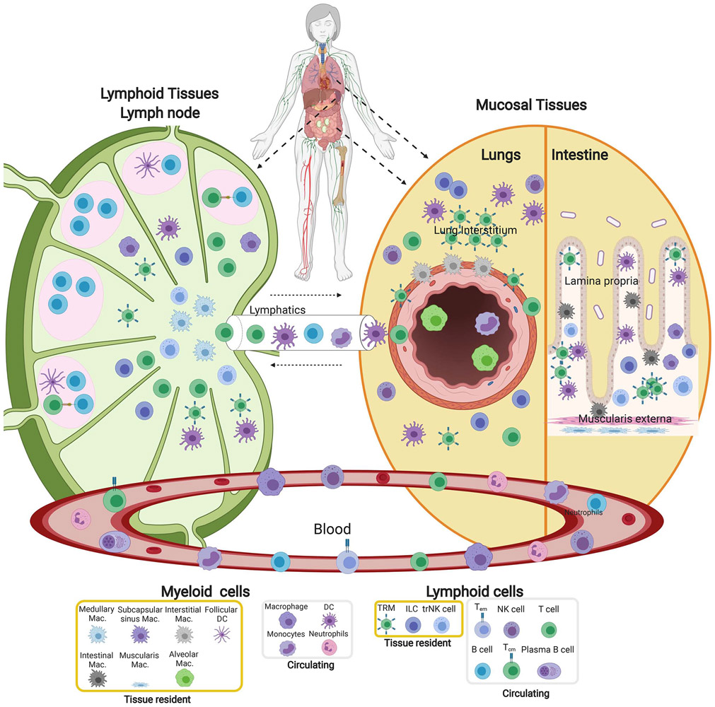
Figure 2: Compartmentalization of immune cells in blood and tissue sites.
The immune system is organized in multiple tissue sites throughout the body; the distribution and composition of immune cells are specific to the tissue sites. Diagram depicts the structural organization and composition of immune cells in mucosal sites (right, lungs and intestines), a lymph node as a representative secondary lymphoid organ (left) and their distinct localization relative to the major epithelial cells and structures of each site. Mucosal sites contain macrophages and tissue resident T cells in the epithelial layers (interstitium) of airways and intestinal villi. In the lymph node, B cells are situated in follicles along with DC surrounded by T cell areas and distinct macrophage subsets in T cell areas. Germinal centers within follicles are the sites of T-B cell interactions and differentiation of B cells to antibody-secreting cells. Also depicted are the major conduits to circulation (Blood, lymphatics) and circulating immune cells. The identity of individual cell subsets is indicated in the legend at bottom.

