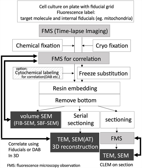
Fig. 1.

Workflow of the live CLEM combined with light microscopic time-lapse imaging and EM. Fluorescence microscope imaging process using wide-field microscope, confocal microscopy, multiphoton microscopy, and types of super-resolution microscopies. Dark boxed area indicating correlative observation process using EM. Volume SEM, including FIB-SEM and SBF-SEM, which are automated 3D reconstruction methods.
