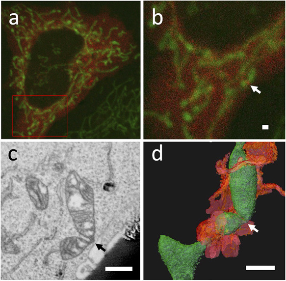
Fig. 2.

Live 3D CLEM of punctuate of DRP1 in HeLa cell. The cells were labeled with PDHA1-GFP for mitochondria (green) and mCherry-DRP1 (red) (a, b). Punctuate of DRP1 was observed on mitochondria (b arrow). Virtual section obtained from a 3D reconstructed dataset from FIB-SEM tomography data (c), and a 3D view of the dataset from the same direction as the optical microscope (d). The arrow in (c) and (d) correspond to the same locations as the arrow in (b). Green-colored objects denote the mitochondria, and orange-colored objects indicate the surrounding endoplasmic reticulum in d. bar = 1 μm.
