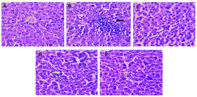
Figure 2.
- Photomicrographs of rat liver tissue (40x). A) Liver tissue in the normal control shows normal hepatocytes with intact portal tracts and central vein; portal tracts show normal portal triad with bile duct and hepatic artery. B) Whereas, the diabetic control rats liver tissue revealed derangement of cells around the central vein, destructed portal triad and central vein, spotty necrosis, ballooning degeneration, apoptosis, portal triditis and piece meal necrosis (black arrow), Kupffer cell hyperplasia (white arrow), sinusoidal and venous congestion. C) The pioglitazone (10 mg/kg body weight), D) and caffeine (20 mg/kg body weight), treated rats reversed the cellular dearrangement around the central vein, and decreased the necrosis. E) Rats treated with pioglitazone + caffeine revealed significant protection of liver with normal microvasculature along with normal hepatocytes.

