Fig. 2.
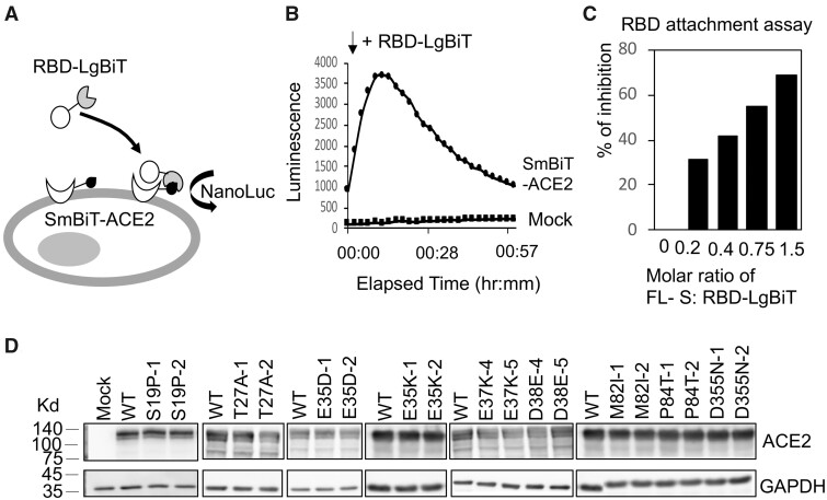
The interaction between RBD and ACE2 variants measured by the RBD attachment assay. (A) Schematic illustration of the RBD attachment assay. Complementation of an active NanoLuc is achieved when the LgBiT and Sm-BiT are brought together upon interaction of RBD and ACE2. (B) HeLa cells transfected with mock construct or SmBiT-ACE2 were treated with RBD-LgBiT and subjected to bioluminescence detection for 1 h. Peak bioluminescence signal was detected at 10 min after the addition of RBD-LgBiT. (C) FLS was mixed with RBD-LgBiT at different molar ratios before incubation with HeLa cells transfected with SmBiT-ACE2. Bioluminescence signal was reduced by increasing the molar ratio of FLS. (D) Expression levels of the wild-type and human ACE2 variants were confirmed by western blotting. Two separate clones were tested for each variant transfection. The GAPDH expression served as an internal control for each experiment.

