
Fig. 3.
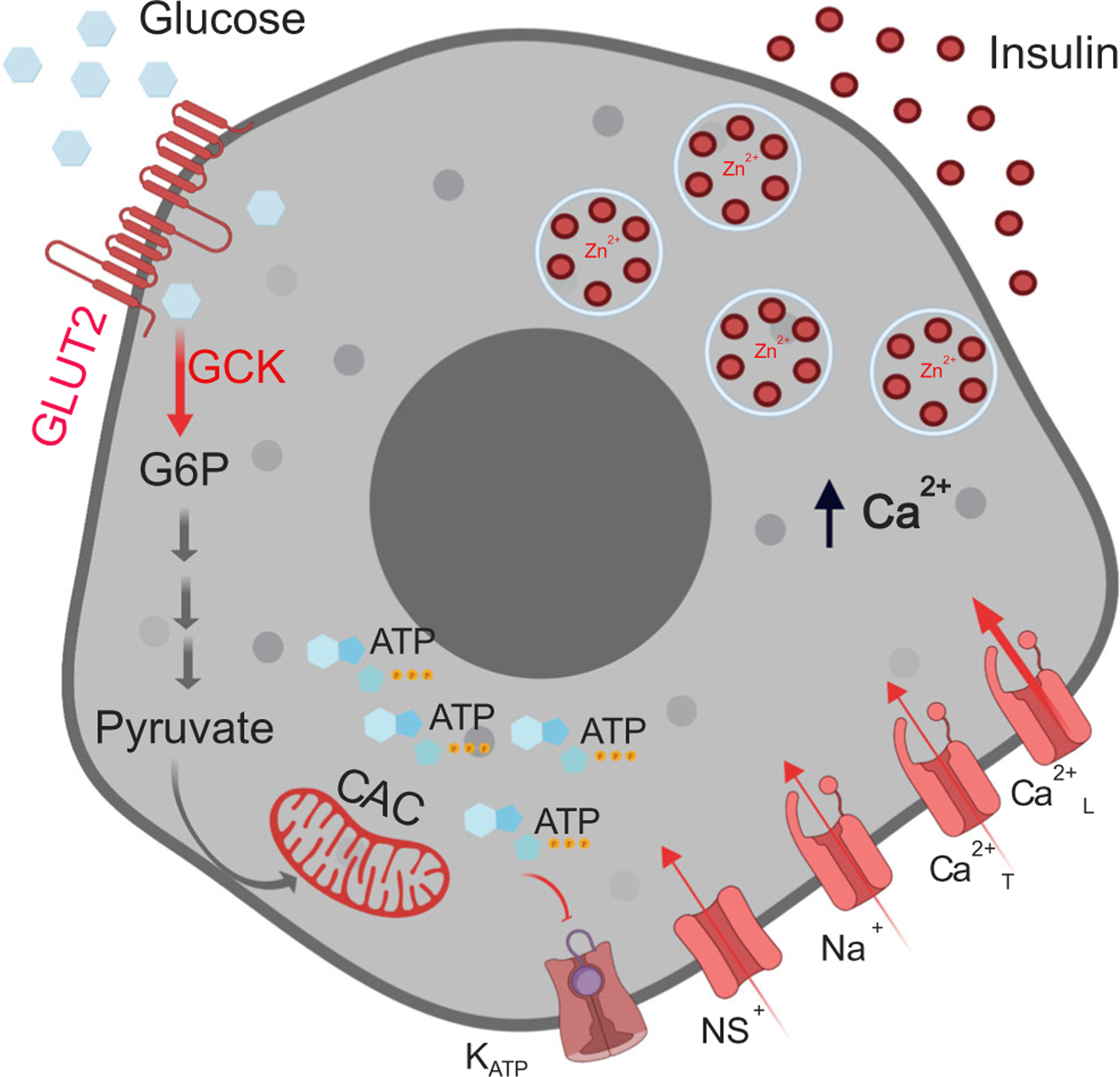
Glucose stimulus–insulin secretion coupling mechanism in beta cells. As extracellular glucose concentrations increase, glucose uptake via GLUT2 increases glucose metabolism via glycolysis (glucokinase-GCK), the citric acid cycle (CAC) and oxidative phosphorylation, increasing ATP production. This, in turn, leads to closure of the KATP channels, which, along with continued conductance through non-selective cationic channels (NS+), results in membrane depolarisation. When the membrane potential reaches a certain threshold, voltage-gated sodium (Na+) channels and T-type Ca2+ channels (Ca2+T) open increasing the membrane potential further to activate the L-type Ca2+ channels (Ca2+L). The associated Ca2+ influx triggers exocytosis of insulin-containing secretory granules. Shown in the different shades of red are components decreased in aged and senescent cells that could result in decreased insulin secretion. G6P, glucose 6-phosphate. Created in BioRender.com.