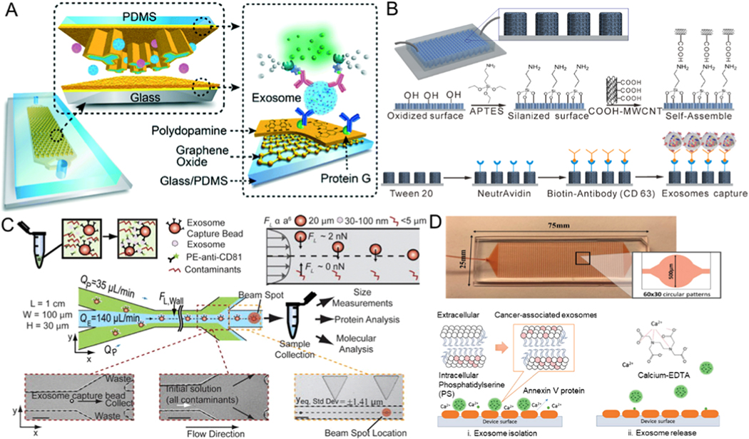
Figure 2.
Microfluidic exosome isolation based on immunoaffinity. (A) Capturing of exosomes on a GO/PDA nanoroughness interface in the nano-IMEX platform. Reprinted with permission from Ref. [50]. Copyright 2016, Royal Society of Chemistry. (B) Schematic illustration of the process of chemical modification with anti-CD63 antibody on 3D MWCNTs functionalized PDMS micropillars for immunocapturing of exosomes. Reprinted with permission from Ref. [52]. Copyright 2017, American Chemical Society. (C) Exosomes captured using magnetic beads and combining the complex with inertial lift forces in a microfluidic channel to further purify the isolated exosomes from other cellular contaminants. Reprinted with permission from Ref. [55]. Copyright 2015, American Institute of Physics. (D) 3D ripple-like structure chip for the immunocapture and release of exosomes based on the interaction between annexin V and phosphatidylserine. Reprinted with permission from Ref. [57]. Copyright 2019,Wiley.

