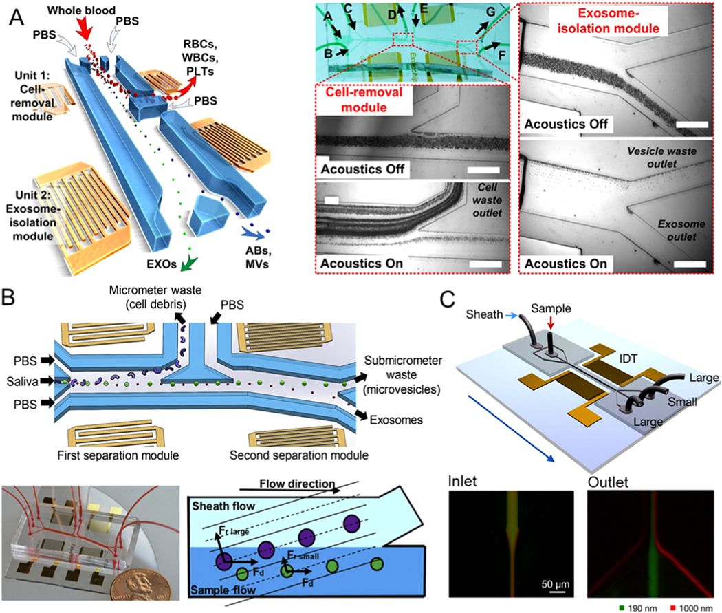
Figure 5.

Microfluidic exosome isolation based on the acoustic field. (A) Schematic of the microfluidic device integrated with acoustic mode for label-free isolation of exosomes. Poly(oxyethylene) as a sheath fluid is introduced from inlet II, while the sample is introduced from inlet I. Exosomes are collected at the side outlet, while larger vesicles are flowing to the middle outlet. Reprinted with permission from Ref. [92]. Copyright 2017, National Academy of Sciences. (B) Schematic and optical image of the acoustic fluidic device for salivary exosome separation. Poly(oxyethylene) as a sheath fluid is introduced from inlet II, while the sample is introduced from inlet I. Exosomes are collected at the side outlet, while larger vesicles are flowing to the middle outlet. Reprinted with permission from Ref. [93]. Copyright 2020, Elsevier. (C) Illustration of the microfluidic device combined with acoustic nano-filter to label-free and size-specifically isolate exosomes. The standing acoustic waves are generated by the interdigitated transducer electrodes. Larger microvesicles are collected at the two side outlets, while smaller exosomes are collected at the middle outlets. Reprinted with permission from Ref. [94]. Copyright 2015, American Chemical Society.
