Fig 1.

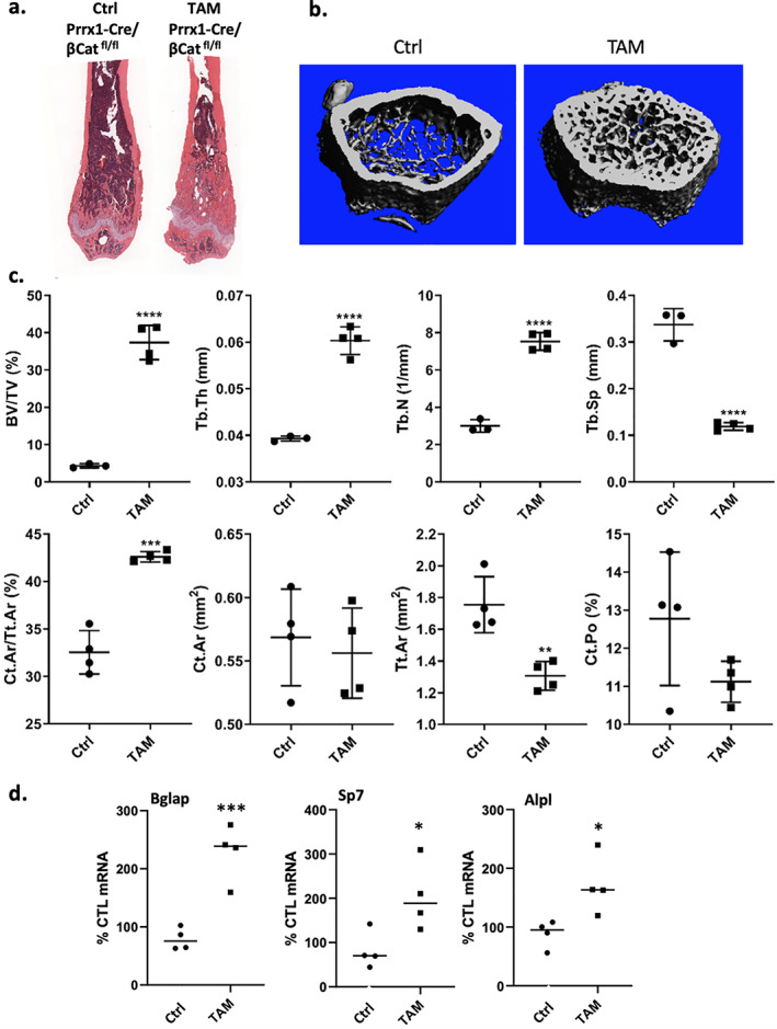
Bone parameters significantly increased after TAM treatment in Prrx1‐Cre/βCatfl/fl mice. Female mice (n = 5 group) were injected with vehicle or TAM (100 μL/mouse, 30 mg/kg × 5; total dose 150 mg/kg dosed over 10 days) beginning at 4 weeks of age and bones harvested at 8 weeks. (A) Paraffin‐embedded H&E‐stained tibia sections. (B) μCT reconstructions of femurs demonstrate more bone in TAM versus VEH; each point represents a separate mouse. (C) Femoral μCT parameters for trabecular and cortical sites. (D) Real‐time PCR for bone formation marker genes with RNA from tibial bone (±SE). Animal data is presented as means ± SD; statistical significance indicated on plots: *p < .05, **p < 0.01, ***p < 0.001 and ****p < 0.0001.
