
Fig. 1.
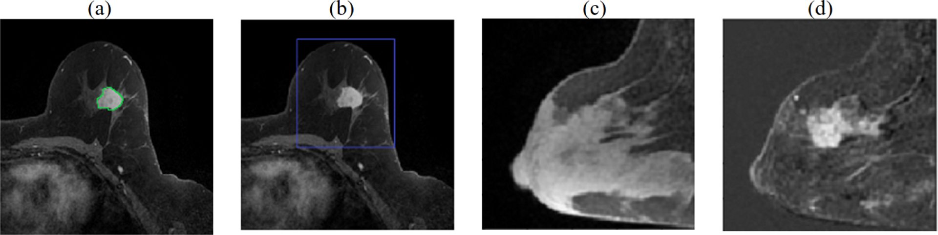
Preprocessing the volumetric DCE MRI. (a) primary tumor is radiologist delineated at time3 in each slice (green contour), (b) MRI is cropped to a cuboidal volume around tumor, (c) sagittal view showing breast at time1, (d) tumor is enhanced by computing difference images, shown here: time3-time1.