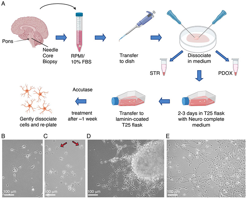
Figure 2: Establishment of tissue culture models from biopsy.
(A) Diagram of general workflow for the establishment of tissue culture models from biopsy. Representative images of (B) early adherent tumor cells, (C) macrophages (indicated by arrows), (D) migratory tumor cells, and (E) tumor cell monolayer from tissue culture establishment of PBT-29FH.

