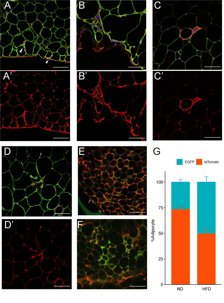
Fig 3. Adipose tissue lineage tracing.
Meflin-CreERT2/Rosa26-tdTomato mice were fed a normal diet for 4 weeks (ND 4w) or a high-fat diet for 8 weeks (HFD 8w) and gonadal WAT (gWAT) samples were examined. WAT was stained with the adipocyte markers perilipin (green), tdTomato (red), and DAPI (blue). (A) ND4w gWAT is surrounded by membranous structures with tdTomato signals. Meflin-expressing lineage cells appear to have penetrated into the adipose tissue stroma (arrowhead). (B) Strong magnified image of membranous structure in ND4w gWAT. (C) HFD8w gWAT. Meflin-expressing lineage cells comprise Crown-like structure, a characteristic structure found in adipose tissue. (D) HFD8w gWAT. Small, multiporous adipocytes are visible, and tdTomato-positive cells can be seen adjacent to the cells. MeflinCreERT2/ROSAmTmG mice were loaded with a normal diet (E) and a high-fat diet (F) for 8 weeks. Whole-mounted gWAT samples were then examined. Confocal laser microscopy was used to image tdTomato-positive and EGFP-positive adipocytes. Each fluorescent cell was counted (G). Scale bars represent 50 μm in (B) and (B’) and 100 μm in all the other panels. A-D: Merged (tdTomato + Perilipin + DAPI), A’-D’: tdTomato, E-F: EGFP + tdTomato.

