
Fig. 1. Preparation and characterization of CHI-C sponge and observation of CHI-C/blood (and plasma) membrane formation named the blood protein barrier.
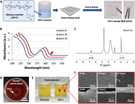
(A) Chemical structure of CHI-C and schematic illustration of the preparation of the CHI-C sponge. Photo credit: Keumyeon Kim, KAIST and InnoTherapy Inc. (B) UV-vis and (C) 1H-NMR spectra of CHI-C. (D) A photographic image of the blood protein barrier (BpB) membrane formed by CHI-C in human whole blood. Photo credit: Keumyeon Kim, KAIST and InnoTherapy Inc. (E) A photographic image of BpB membranes (right vial, red arrows) formed by CHI-C in human plasma. However, no BpB membrane was formed by chemically unmodified chitosan (left vial). Photo credit: Keumyeon Kim, KAIST and InnoTherapy Inc. (F) Scanning electron microscope (SEM) images of the surface (top) and cross section (bottom) of the BpB membrane at predetermined immersion times (3 min, 3 hours, and 12 hours). a.u., absorbance unit.