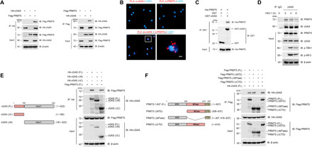
Fig. 1. PRMT5 interacts with cGAS.

(A) Co-IP analysis of the interaction between PRMT5 and cGAS in HEK293T cells cotransfected with cGAS and PRMT5 plasmids. IP assay was done by anti-HA (left) and anti-Flag (left) antibodies to show the interaction between PRMT5 and cGAS. (B) In situ PLA analysis of the direct interaction of PRMT5 with cGAS in mouse macrophage. Scale bars, 10 μm. (C) GST pull-down analysis of purified PRMT5 incubated with GST or GST-cGAS in vitro. (D) IP analysis of the interaction between endogenous PRMT5 and cGAS in mouse BMDM cells infected with HSV-1 [multiplicity of infection (MOI) = 1] for 0, 3, and 6 hours. (E and F) Full-length (FL) and truncated mutant schematic structures of cGAS (E) and PRMT5 (F) were presented (left), and co-IP assay was performed to analyze the interaction between the full-length or truncated mutant forms of PRMT5 and cGAS (right). Data are representative of three independent experiments with similar results.
