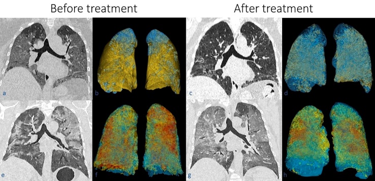
Figure 4.
The images of the first row correspond to patient 1 (a, b, c, d), while images of the second row correspond to patient 4 (e, f, g, and h). CT scans in coronal plane are shown before (a, e) and after treatment (c, g) with their respective three-dimensional segmentation (b, and f before treatment; c and g after treatment). Notice in the affected lung parenchyma in the CT scans as ground-glass opacities and consolidations. In the three-dimensional lung segmentation, blue corresponds to undamaged parenchyma, the yellow color corresponds to ground-glass opacities, while the red color corresponds to consolidation. Notice the reduction of ground-glass opacities and consolidations in the lungs of both patients after treatment.

