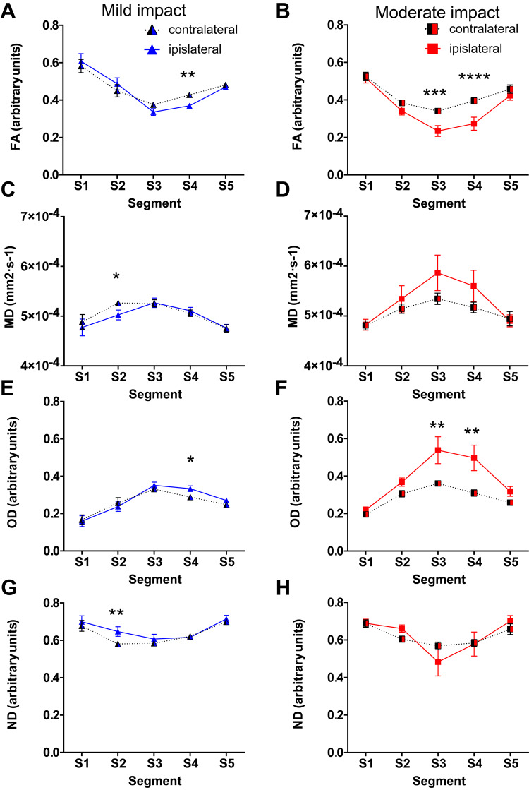
Figure 4.
Diffusion tensor imaging measures show white matter damage in corpus callosum segments subjected to highest strain. Diffusion tensor imaging measures in segments of the corpus callosum across the ipsilateral and contralateral hemispheres. Fourteen days after mild/moderate impact, mean values of: (A and B) FA; (C and D) MD; (E and F) Orientation dispersion (OD); and (G and H) Neurite density (ND). All data are presented as mean ± SEM. Mild impact: n = 10, moderate CCI: n = 11. *P < 0.05, **P < 0.01, ***P < 0.001 and ****P < 0.0001 as compared to the contralateral side.

