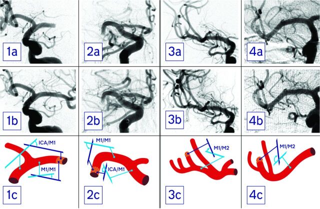
Fig 1.
First and second columns (patients 1 and 2) show pre- (a) and postprocedural (b) angiograms of patients with a proximal M1 occlusion as well as schematics of the vessel anatomy (c). The thrombus site is hatched red and yellow. In patient 1, the ICA/M1 was 53° and the M1/M1 was 18°, and he was successfully recanalized (TICI 3). Patient 2 presented with an ICA/M1 of 140° and an M1/M1 of 105°. In this patient, thrombectomy was unsuccessful (TICI 0). Third and fourth columns (patients 3 and 4) show pre- (a) and postprocedural (b) angiograms of patients with a proximal M2 occlusion as well as vessel schematics (c). While patient 3 presented with an M1/M2 angle of 51° and was successfully recanalized (TICI 3), in patient 4, an M1/M2 angle of 128° was measured and thrombectomy was unsuccessful (TICI 1).

