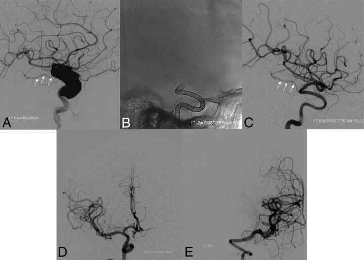
Fig 1.
Baseline pretreatment digital subtraction angiogram (lateral projection, A) demonstrating a large dysplastic ICA aneurysm with preserved antegrade flow in the anterior choroidal artery (white arrows). In an initial posttreatment follow-up angiogram after placement of a PED, unsubtracted mask (B) and arterial phase DSA in a lateral projection (C) demonstrate complete occlusion of the aneurysm with preservation of antegrade flow within the anterior choroidal artery (white arrows). Follow-up DSAs demonstrate reconstruction of the left ICA with smooth uniform neointimal overgrowth of the minimally porous endoluminal device construct. Eighteen-month follow-up DSAs (frontal projections) demonstrate a contralateral supply of the left anterior cerebral artery from the right ICA (D) and persistent occlusion of the left ICA aneurysm (E).

