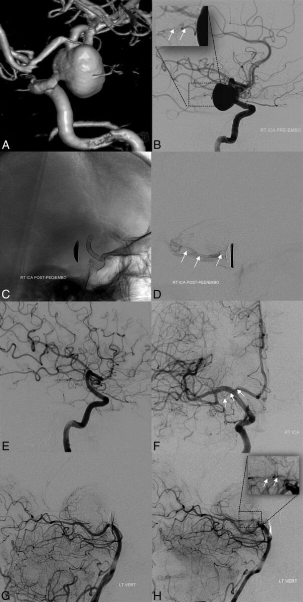
Fig 3.

Baseline pretreatment 3D-DSA (A) and lateral projection DSA (B) demonstrating a large anterior choroidal artery aneurysm. Note the origin of the AchoA from the aneurysm fundus (white arrows in inset, B). Immediate posttreatment images, after placement of PED, unsubtracted mask (C) and delayed image DSA (D), demonstrate coverage of the AchoA origin and residual antegrade opacification of the AchoA territory (white arrows, D). Follow-up angiography (E and F) confirms complete occlusion of the aneurysm and AchoA origin; the AchoA is opacified retrogradely (white arrows, F). Baseline pretreatment (G) and follow-up (H) DSAs of the left vertebral artery (lateral projections) demonstrate the collateral opacification of the AchoA through anastomoses with the posterior lateral choroidal artery at follow-up (inset in H, white arrows; compare with the inset in B), not visualized at baseline (G).
