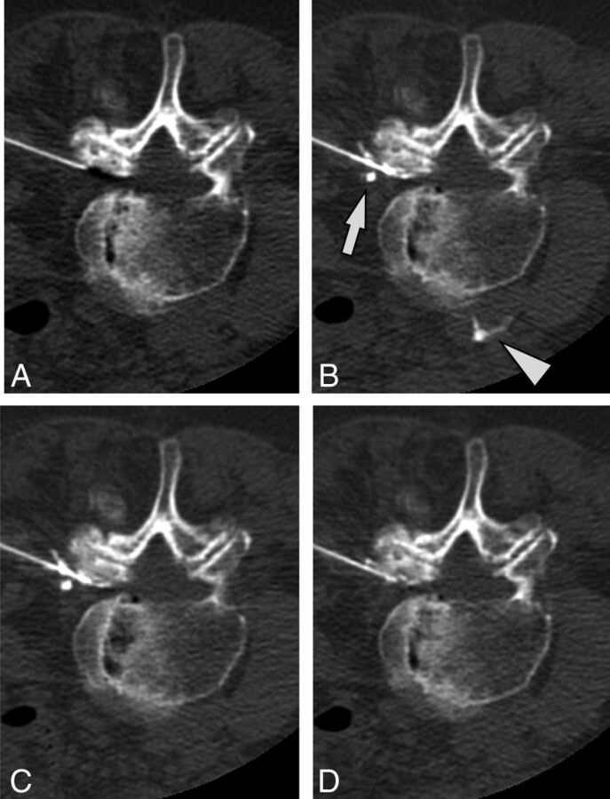
Fig 3.
A case of intravascular injection is demonstrated on preinjection (A), immediate postinjection (B), first delayed (C) images, and an additional delayed (D) image. On the immediate postinjection image, contrast is seen in the ascending lumbar vein (arrow) and the inferior vena cava (arrowhead). There is washout of contrast from the inferior vena cava on the first delayed image and from the ascending lumbar vein on the second delayed image.

