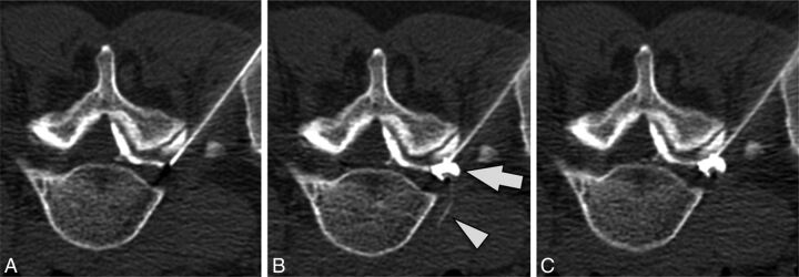
Fig 8.
Simultaneous epidural and intravascular injection. Preinjection (A), immediate postinjection (B), and delayed postinjection (C) images demonstrate a vessel extending into the anterior paraspinal soft tissues (arrowhead) that washes out on the delayed image. Epidural contrast in the neural foramen (arrow) persists on the delayed image. Note that there is contrast in the epidural space of the spinal canal on the preinjection image due to prior injection at an adjacent level.

