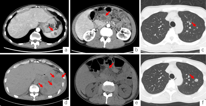
Figure 2.
Axial contrast-enhanced computed tomography shows splenic hypodense abscess (arrow) (a), enlarged mesenteric lymph nodes (arrow) (b), and a lung nodule in the right upper lobe (arrow) (c). For 10 consecutive days, the splenic lesions increased in size and number (arrows) (d); however, the mesenteric lymph nodes showed no changes (arrow) (e), and the lung nodule increased in size and formed a cavity (arrow) (f).

