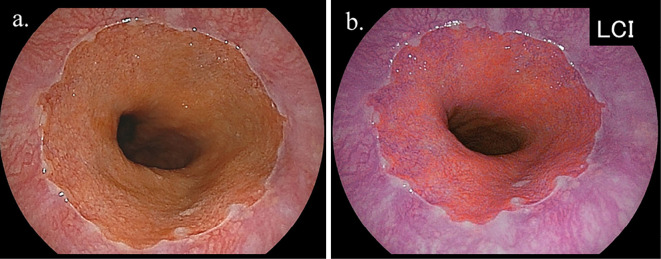
Figure 3.
Representative endoscopic findings of short segment Barrett’s esophagus (SSBE) in subject without H. pylori infection [a: white light imaging (WLI), b: linked color imaging (LCI)]. The presence of palisade vessels in the area of columnar lined epithelium was easily diagnosed by LCI, although it was not easily recognized by WLI. The SSBE length was classified as <10 mm in this case.

