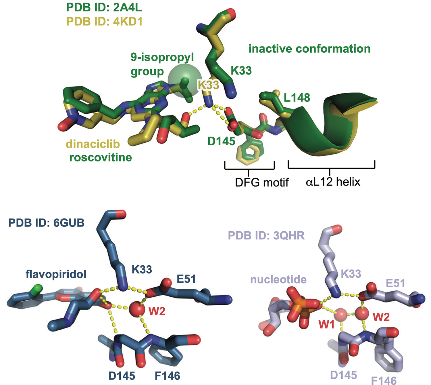
Extended Data Fig. 1. Cdk2 inhibitors drive conformational shifts upon binding.

Aligned crystal structures of Cdk2 bound to dinaciclib and roscovitine (top), and structures of Cdk2:cyclinA bound to flavopiridol and ADP shown side by side (bottom). Hydrogen bonds are shown as yellow dashed lines, and structured water molecules as red spheres. The structure of Cdk2 bound to AZD5438 is shown in Supplementary Fig. 7.
